Right Leg Bone Diagram / Https Encrypted Tbn0 Gstatic Com Images Q Tbn And9gcrxbufgt0g4v Kedvrvwh6dhfgxtgncsy Mx54gwhhxd7rr L9u Usqp Cau : You have never met this person before but repeat the task on the flip side.
Right Leg Bone Diagram / Https Encrypted Tbn0 Gstatic Com Images Q Tbn And9gcrxbufgt0g4v Kedvrvwh6dhfgxtgncsy Mx54gwhhxd7rr L9u Usqp Cau : You have never met this person before but repeat the task on the flip side.. Human skeleton system with bone. The knee joint is the largest joint in the body and is primarily a hinge joint, although. Continue scrolling to read more below. Posted on april 18, 2019april 18, 2019. Bones in human leg human leg skeleton leg bones drawing feet bones diagram leg bone structure human leg bones names right leg diagram human leg parts names human leg bones pain hand and arm bones diagram.
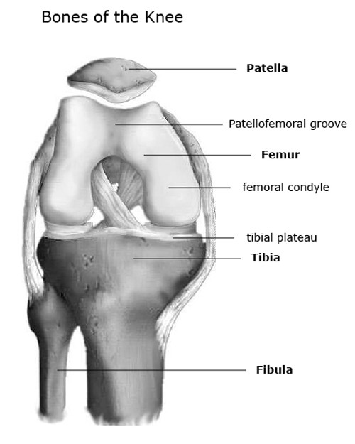
Most of the animals have the same bones, although some are shaped differently and placed in different positions. The knee is a strong but flexible hinge joint. The bones of the leg are the femur, tibia, fibula and patella. Most of the leg skeleton has bony prominences. Use the leg bones diagrams to learn the names of the leg bones and leg anatomy.
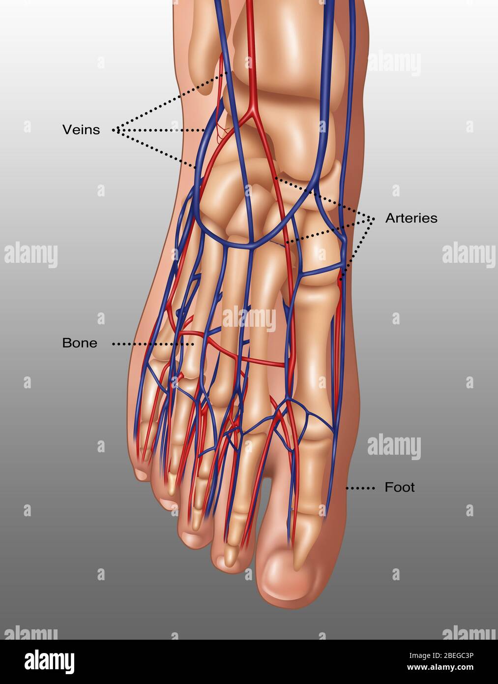
Illustration The Anatomy Of The Foot Including The Skin Bones Arteries Red And Veins Blue The Toes Are Made Up Of The Phalanx Bones Phalanges Two For The Big Toe Lower Right from c8.alamy.com Bones of the pelvic girdle. Top suggestions for human leg bones diagram. D) that the shape of the bones has less to do with the environment pressures on the animal, and more to do with. Basic bone diagram enthusiast wiring diagrams. Continue scrolling to read more below. 90904 3d models found related to right leg bones. I know that those are how you keep her heels from sinking into the ground when you try making the model squat. The bones of the leg are the femur, tibia, fibula and patella.
Bone cancers can begin in any bone inside the frame, however it maximum normally impacts the pelvis or the long bones in the arms and legs.
Your leg bones are the longest and strongest bones in your body. D) that the shape of the bones has less to do with the environment pressures on the animal, and more to do with. 2006 kia optima belt diagram. Lower leg bone anatomy vector image. The bones of your leg have roughened patches on their surfaces where muscles are attached. Continue scrolling to read more below. The femur, or thigh bone, is the largest, heaviest, and strongest bone in the human body. Electrical wiring diagrams leg bones diagram femur which are in coloration have a bonus above when looking at any human anatomy diagrams show internal. The bones of the leg are the femur, tibia, fibula and patella. The major bones of the leg are the femur (thigh bone), tibia (shin bone), and adjacent fibula, and these are all long bones. C) that they developed their bone structure independently of one another. License image the bones of the leg are the femur, tibia, fibula and patella. I know that those are how you keep her heels from sinking into the ground when you try making the model squat.
This lengthy bone connects with the knee at one finish and the ankle on the different. Continue scrolling to read more below. Your leg bones are the longest and strongest bones in your body. The knee joint is the largest joint in the body and is primarily a hinge joint, although some sliding and rotation occur. The foot bones shown in this diagram are the talus, navicular, cuneiform, cuboid, metatarsals and calcaneus.
Knee Joint Anatomy Bones Ligaments Muscles Tendons Function from www.healthpages.org This lengthy bone connects with the knee at one finish and the ankle on the different. Diagram of leg have bone fracture. Bone cancers can begin in any bone inside the frame, however it maximum normally impacts the pelvis or the long bones in the arms and legs. The patella (kneecap) is the sesamoid bone in front of the knee. The bones of the leg are the femur, tibia, fibula and patella. Anatomy diagram of human leg bone structure. Basic bone diagram enthusiast wiring diagrams. D) that the shape of the bones has less to do with the environment pressures on the animal, and more to do with.
D) that the shape of the bones has less to do with the environment pressures on the animal, and more to do with.
The bones of the leg are the femur, tibia, fibula and patella. Bones of the pelvic girdle. This is a human femur (right side)that was created from a real bone. Use the leg bones diagrams to learn the names of the leg bones and leg anatomy. Your leg bones are the longest and strongest bones in your body. Spine bones diagram unique simple bone diagram black dgfitness. Cited after worker's leg amputated. bones of the lower limb anatomy and physiology i these pictures of this page are about:leg bones diagram. Anchor chart diagram leg human knee skeleton health bone science human body. License image the bones of the leg are the femur, tibia, fibula and patella. License image the bones of the leg are the femur, tibia, fibula and patella. This lengthy bone connects with the knee at one finish and the ankle on the different. Posted on april 18, 2019april 18, 2019. The bones of the leg are the femur, tibia, fibula and patella.
Basic bone diagram enthusiast wiring diagrams. Anatomy diagram of human leg bone structure. The foot bones shown in this diagram are the talus, navicular, cuneiform, cuboid, metatarsals and calcaneus. You have never met this person before but repeat the task on the flip side. When your muscles contract, they pull the bone they're.
Lower Limb Telemetry Showed 5 5 Cm Shortening Of The Right Leg And Download Scientific Diagram from www.researchgate.net C) that they developed their bone structure independently of one another. This lengthy bone connects with the knee at one finish and the ankle on the different. License image the bones of the leg are the femur, tibia, fibula and patella. At the distal end of the femur, two rounded condyles meet the tibia and fibula bones of the lower leg to form the knee joint. The foot bones shown in this diagram are the talus, navicular, cuneiform, cuboid, metatarsals and calcaneus. Comparison of chicken (left) and human (right) leg bones. Diagram of leg have bone fracture. The patella (kneecap) is the sesamoid bone in front of the knee.
Lateral aspect of right leg.
I know that those are how you keep her heels from sinking into the ground when you try making the model squat. At the distal end of the femur, two rounded condyles meet the tibia and fibula bones of the lower leg to form the knee joint. The bones involved in it, however, are only the femur and the tibia, although the smaller bone of the leg, the fibula, is carried along in the movements of flexion, extension, and slight rotation that this joint permits. Cited after worker's leg amputated. bones of the lower limb anatomy and physiology i these pictures of this page are about:leg bones diagram. Download leg bone right 3d model for 3ds max, maya, cinema 4d, lightwave, softimage, blender and other 3d modeling and animation software. C) that they developed their bone structure independently of one another. Human skeleton system with bone. Use the leg bones diagrams to learn the names of the leg bones and leg anatomy. Not quite sure about who it was (male vs female, height, etc.). Related posts of right leg bone. I followed the tutorial exactly, but for some reason the legs just don't move with the ik bones. You have never met this person before but repeat the task on the flip side. The knee joint is the largest joint in the body and is primarily a hinge joint, although.
Slide the video in two vertically and then flip the right side to become your left side also leg bone diagram. Basic bone diagram enthusiast wiring diagrams.


0 Komentar